Sunday 23 June 2024

Lethal & Conditional Mutation, LOF, GOF, Gain of function & loss of function

 Content

  • Introduction 
  • Origin 
  • What is a gene mutation and how do mutations occur? 
  • How the gene mutation affect heath and Development?  
  • Lethal & Mutation  
  • LOF And GOF Mutation 
  • Software Tools for Genetic Mutation Analysis 
  • Reference
Origin

Meaning: The term was derived from a Greek word 'Muto' which means 'to change. Therefore, mutation may be defined as "any change in the genetic material of organisms which is permanent and heritable from generation to generation

Definition: Process by which sequences of base pairs is altered. Mutation could result in changes in base pairs and in chromosome. The term mutation was given by Hugo de Vries(1901).

What is a gene mutation and how do mutations occur?

  • Gene mutation are changes in the sequence of a DNA.
  • Gene mutations can be classified in two major ways: 
    • Hereditary (Germinal):- Germline mutation occur in a parent’s reproductive cell (egg or Sperm). These mutation change the genetic material that the child receives from their parent. You can inherit germline mutation from either parent. 
    •  Aquired (Somatic):- Somatic mutation are a change to a person’s DNA that occurs after conception to any cell that is not germ cell. Somatic mutations don’t pass from parent’s to their child and happen randomly without the mutation existing in a person’s family history. They also can’t pass to future generations.
How can gene mutations affect health and development?

  • To function correctly, each cell depends on thousands of proteins to do their jobs in the right places at the right times. 
  • Sometimes, gene variants prevent one or more proteins from working properly. By changing a gene's instructions for making a protein, a variant can cause a protein to not to be produced at all. 
  • When a variant alters a protein that plays a critical role in the body, it can disrupt normal development or cause a health condition.
  • A condition caused by variants in one or more genes is called a genetic disorder. 
  • Gens themselves do not cause disease-Genetic disorders are caused by variants that eliminate a gene's function. Example when people say that someone has "the cystic fibrosis gene" they are usually referring to a version of the CFTR gene that contain a variant that causes the disease. All people including those without cystic fibrosis, have version of the CFTR gene.
Do all gene mutations affect health and development?

No; only a small percentage of mutations cause genetic disorders—most have no impact on health or development. For example, some mutations alter a gene's DNA sequence but do not change the function of the protein made by the gene.

Lucien Claude Marie Julien Cuénot (French: [keno]; 21 October 1866 – 7 January 1951) was a French biologist. In the first half of the 20th century, Mendelism was not a popular subject among French biologists. Cuénot defied popular opinion and shirked the “pseudo-sciences” as he called them. Upon the rediscovery of Mendel's work by CorrensDe Vries, and Tschermak, Cuénot proved that Mendelism applied to animals as well as plants.


Fig,-Lucien Claude


Lethal Mutation
  • Lethal mutation is a type of mutation in which Lethal means that something has an intense enough impact to cause the death of an organism. So a lethal mutation is a kind of mutation in which the effect(s) might cause mortality or greatly lower the predicted lifespan of an organism bearing the mutation.
  • Are all mutations lethal? No, Some mutations may have little to no effect on the organism it impacts. Others mutations may cause disabilities to the organism, whether physically or mentally. This is known as a non-lethal mutation. However, some mutations are quite lethal.
Lethal allele
  • A classic example of an allele that affects survival is the lethal yellow allele, a spontaneous mutation in mice that makes their coats yellow. This allele was discovered around the turn of the 20th century by the French geneticist Lucien Cuenót, who noticed that it was inherited in an unusual pattern.
  • When yellow mice were crossed with normal brown mice, they produced half yellow and half brown offspring. This suggested that the yellow mice were heterozygous, and that the yellow allele, A^Y, was dominant to the brown allele, A. But when two yellow mice were crossed with each other, they produced yellow and brown offspring in a ratio of 2:1, and the yellow offspring did not breed true (were heterozygous). Why was this the case?
  • • As it turned out, this unusual ratio reflected that some of the mouse embryos (homozygous (A^y A^y) genotype) died very early in development, long before birth. In other words, at the level of eggs, sperm, and fertilization, the color gene segregated normally, resulting in embryos with a 1:2:1 ratio of (A^y A^y), (A^y A), and (AA) genotypes. However, the (A^y A^y) mice died as tiny embryos, leaving a 2:1 genotype and phenotype ratio among the surviving mice. 
  • Alleles like A^y, which are lethal when they're homozygous but not when they're heterozygous, are called recessive lethal alleles.

Recessive lethality – 
  • Sickle Cell anemia
  • Cystic Fibrosis 
  • Achondroplasia
Dominant lethality – 
  • Familial Hypercholesteramia 
  • Huntington Disease 
  • Neurofibromatosis
  • Marfan Syndrome
Types of lethal genes

Recessive Lethal Genes 
  • Cuénot and Baur discovered these first recessive lethal genes because they altered Mendelian inheritance ratios. 
  • Recessive lethal genes can code for either dominant or recessive traits, but they do not actually cause death unless an organism carries two copies of the lethal allele. 
  • Examples of human diseases caused by recessive lethal alleles include cystic fibrosis, sickle-cell anemia, and achondroplasia, 
  • Achondroplasia is an autosomal dominant bone disorder that causes dwarfism. While the inheritance of one achondroplasia allele can cause the disease, the inheritance of two recessive lethal alleles is fatal.
Dominant Lethal Genes

  • Dominant lethal genes are expressed in both homozygotes and heterozygotes. But how can alleles like this be passed from one generation to the next if they cause death?
  • Dominant lethal genes are rarely detected due to their rapid elimination from populations. One example of a disease caused by a dominant lethal allele is Huntington's disease. 
  • Huntington's disease
  • A neurological disorder in humans, which reduces life expectancy.
  • Because the onset of Huntington's disease is slow, individuals carrying the allele can pass it on to their offspring. This allows the allele to be maintained in the population. Dominant traits can also be maintained in the population through recurrent mutations.
Conditional Lethal Genes 
  • an organism lives normally under one set of conditions, but when certain changes are introduced in its environment, lethality results. 
  • Favism is a sex-linked, inherited condition that results from deficiency in an enzyme called glucose-6-phosphate dehydrogenase. 
  • It is most common among people of Mediterranean, African, Southeast Asian, and Sephardic Jewish descent.
  • The disease was named because when affected individuals eat fava beans, they develop hemolytic anemia, a condition in which red blood cells break apart and block blood vessels. 
  • Blockage can cause kidney failure and result in death.


Sex-Linked Lethal Genes 
  • Semilethal or Sublethal Genes. 
  • the lethal gene is carried on the sex chromosome, usually X. 
  • Hemophilia is a hereditary disease caused by deficiencies in clotting factors, which results in impaired blood clotting and coagulation.
  • Because the allele responsible for hemophilia is carried on the X chromosome, affected individuals are predominantly males, and they inherit the allele from their mothers. 
  • The alleles responsible for hemophilia are thus called semilethal or sublethal genes, because they cause the death of only some of the individuals or organisms with the affected genotype.
  • Normally, clotting factors help form a temporary scab after a blood vessel is injured to prevent bleeding, but hemophiliacs cannot heal properly after injuries because of their low levels of blood clotting factors. 
  • Therefore, affected individuals bleed for a longer period of time until clotting occurs. This means that normally minor wounds can be fatal in a person with hemophilia.
Synthetic Lethal Genes 
  • Synthetic lethality describes the genetic interaction between two genes. If either gene is mutated by itself, the organism remains viable. The combination of a mutation in both genes is incompatible with viability and results in lethality.
  • Scientists studying the fruit fly observed that pairwise combinations of some mutant alleles were not viable, whereas singly, the same mutant alleles did not cause death (Boone et al., 2007). In other words, some mutations are only lethal when paired with a second mutation. These genes are called synthetic lethal genes.
  • When an allele causes lethality, this is evidence that the gene must have a critical function in an organism.
  • The discoveries of many lethal alleles have provided information on the functions of genes during development. 
  • So scientists used conditional and synthetic lethal alleles to study the physiological functions and relationships of genes under specific conditions.
LOF & GOF Mutation 
Loss Of Function
  • Gene gain and loss are prevalent in the genomes of diverse organisms and contribute to genetic variation. 
  • The importance of gene loss (or pseudogene formation) has been almost entirely ignored for a long time, mainly because pseudogenes may not produce full length proteins and are regarded as putatively non-functional.
  • the ‘‘less is more’’ hypothesis proposes that gene loss may be an adaptive evolutionary process that is beneficial to organisms.
  • Two major mechanisms can cause gene loss: physical removal events (recombination or the mobilization of transposable or viral elements) that lead to the fragment deletion of one or more genes, and deleterious mutations at gene coding regions that cause loss function (LOF) mutations.
  • An LOF mutation was first observed in the 5S DNA gene of Xenopus laevis. This gene could not encode a functional 5S rRNA and was named a pseudogene.
  • Many pseudogenes caused by LOF mutations were reported in Escherichia coli, yeast, mammals and plants.
Fig.-(A) Different kinds of LOF mutations. (B) LOF mutations affect different isoforms and the mechanism of the NMD pathway. The LOF mutation only affects a part of the gene transcript in case 1 and affects all transcripts in case 2. Orange rectangles indicate exons, gray rectangles indicate frameshift sequences of exons or retained intron sequences, black lines between them indicate introns, and red lines indicate LOF mutations caused by loss of a start codon or gain of a premature stop codon.

GENETIC VARIATIONS THAT CAUSE LOF MUTATIONS

  • There are four major genetic variations that can lead to LOF mutation,
  • First, a nonsense SNP may lead to a premature stop codon, producing a truncated protein sequence. For example, in Arabidopsis, FRIGIDA (FRI) alleles with premature stop codons explain a large fraction of flowering-time variation.
  • Second, a SNP that occurs at a canonical splice site may affect splicing. Specifically, a SNP that occurs in a splice donor site causes intron retention in the mRNA, whereas a SNP that occurs in a splice acceptor site removes an exon from the original mRNA. Splice site variations may eventually lead to frameshifts or premature stop codons. In the Arabidopsis relative Capsella rubella, splice site mutations in the FLOWERING LOCUS C gene cause a premature stop codon that promotes flowering.
  • Third, insertion or deletion variants with non-integral multiples of 3 located in the gene coding region can lead to frameshifts by disrupting the full length transcript.
  • Fourth, the loss of an initiation codon can lead to LOF mutations. The loss of transcription start codon (ATG) variations prevents gene transcription if there is no alternative start codon near the mutation. For example, in humans, the loss of the start codon in the FRMD7 gene leads to idiopathic infantile nystagmus disease.
LOF MUTATIONS ARE ABUNDANT IN DIVERSE SPECIES 

  • LOF mutations have been studied in diverse species at the population level. Almost every genome contains many LOF mutations in either heterozygous or homozygous states. Most LOF mutations in a genome tend to be present at low allele frequencies and have a heterozygous status.
  • In humans, an analysis of 7597 genomes identified 17 764 stop-gain variants and 13 915 frameshift variants within 11 369 protein-coding genes.
  • Based on 1432 whole exome sequences from five isolated European populations, 173 homozygous LOF mutations were identified within 167 genes. 
  • Analysis of whole genomes sequencing from 2636 Icelanders and chip-genotype data from 101 584 additional individuals identified 6795 autosomal LOF variants within 4924 gene. 
  • Our recent study of 1071 A. thaliana genomes from worldwide accessions revealed 60 819 LOF variants within 12 907 genes.
FUNCTIONAL EFFECTS OF LOF MUTATIONS 
  • Compared with functional protein-coding genes (Figure ), LOF mutations are natural gene knockouts that can provide insight into gene function in diverse organisms, 
  • In rice, a natural LOF mutation in GSE5, which encodes a plasma membrane-associated protein, contributes to grain size diversity. 
  • In A. thaliana, natural brx LOF alleles confer root adaptation to acidic soil, and a natural knockout allele of ARMADILLO REPEATCONTAINING KINESIN1 causes root hair branching, 
  • In addition, different LOF mutations of duplicated genes in diverse A. thaliana populations can lead to hybrid incompatibility,
Fig.-(A) Functional genes without LOF mutation. (B) Gene knockout with LOF mutations. (C) Gene translated into functional protein if LOF mutations are located at 30 or 50 termini. (D) Transcriptional readthrough of the premature stop codon. (E) Gene translated to a new functional protein with the LOF mutation. Orange rectangles indicate exons, black lines between them indicate introns, red lines indicate LOF mutations, blue rectangles indicate the core functional domain of the gene, and gray rectangle indicates frameshift. Orange lines with polyadenylic acid indicate mRNA.

ESSENTIAL GENES IN NATURAL POPULATIONS
  • Essential genes are those genes that are necessary for survival. 
  • An individual cannot survive if LOF mutations have occurred in essential genes; otherwise, these genes would be considered non-essential. 
  • LOF mutations in the genomes of natural populations provide direct evidence to delimit essential genes. 
  • Our essential gene number is much larger but understandable. Because growth conditions in laboratories are much better than in natural habitats, plants probably tolerate more LOF mutations when grown in laboratories. Therefore, the definition of essential genes is context dependent, reflecting the niche that the organisms inhabit.
Fig.-The Identification of Essential Genes through LOF Mutations. (A) The identification of single-copy essential genes. The individuals could not survive when LOF mutations occurred in essential genes. (B) The identification of multiple copies of essential genes. When duplication events occurred in essential genes, either one of the copies would be redundant. The individual could survive if LOF mutations occurred in only one of the copies.

LOF MUTATIONS ARE CRUCIAL FOR ADAPTATION AND DIVERSIFICATION 
  • LOF mutations in the genome may be neutral, deleterious, or advantageous. Neutral or less-deleterious LOF mutations can be tolerated and may even accumulate during range expansion. 
  • the ‘‘less is more’’ hypothesis proposes that gene loss may be beneficial to organisms. Adaptive LOF mutations have been observed frequently in bacteria and yeast. An analysis of bacterial fitness in more than 100 different conditions revealed that LOF mutations can provide fitness benefits. 
  • LOF mutations play important roles in the evolution and diversification of diverse organisms. In plants, a premature stop codon in GL4 caused smaller grain size and loss of seed shattering during African rice domestication.
  • Similarly, flower color plays an important role in pollinator attraction (Bradshaw and Schemske, 2003). In Petunia axillaris, an LOF mutation in ANTHOCYANIN2 (AN2) occurred independently at least five times, changing the flower color from violet-red to white compared with Petunia integrifolia and influencing the shift in pollinator attraction from bees to hawkmoths.
  • LOF mutations have also been found to be beneficial in human evolution. A CASP12 LOF allele is known to promote resistance to severe sepsis, and rare LOF mutations in SLC30A8 can protect against type 2 diabetes.
Fig.-Adaptive Evolution of LOF Mutations. LOF mutations can be neutral, deleterious, or advantageous. Deleterious LOF mutations are usually present at low allele frequencies in natural populations due to purifying selection. Neutral or beneficial LOF mutations can be tolerated or may accumulate during range expansion, and beneficial LOF mutations may be fixed by positive selection.

FUTURE DIRECTIONS 
  • Species-wide studies could be performed to understand the genome-wide distribution patterns, functional effects, and evolutionary importance of LOF mutations. Several studies of LOF mutations at the genome level have been performed in natural populations. 
  • In particular, our recent study revealed that the level of nucleotide diversity, the density of transposable elements, and gene family size are positively correlated with the presence of LOF mutations. 
  • the evolutionary effects of natural LOF mutations on other genes in the same pathway may be complicated. When genes become non-functional, other genes in the same pathway may accumulate LOF mutations as well. 
  • the functional effects of LOF mutations are interesting to study. Genome-wide study of LOF variants can provide information on gene lethality based on the frequency of LOF alleles for a given gene in natural environments.
  • In yeast S. cerevisiae, a study of 1106 essential gene knockouts found that 88 (9%) of them could survive through adaptive evolution, and these were defined as evolvable essential genes. 
  • Synthetic biology can offer interesting insights into the beneficial effects of regulator loss. For example, in Chinese hamster ovary cells, repressor loss in the promoter region of PuroR leads to high gene expression and drug resistance.
  • the agricultural importance of LOF mutations is largely unknown. In plants, several studies have reported that LOF mutations can also increase crop yield. For example, LOF of GW2, a gene that encodes a RING-type E3 ubiquitin ligase, can increase grain width and weight. 
  • Similarly, an LOF mutation of MEI2-LIKE PROTEIN4 (OML4) leads to large and heavy grains in rice (Oryza sativa).
Gain of function
  • Gain-of-function mutations-cause either new function or function expressed at new times or location within organism.
  • Gain of function mutation is dominant. Single mutation could derive cell toward cancer. 
Fig.-Ptd1 competitively binds hydrophobic molecules, which may be related to environmental adaptation of rice, resulting in a LD/LT-sensitive growth inhibition phenotype. Through signal transduction, the plant senses a change in the environment and responds to it. Many hydrophobic molecules are involved in environmental adaptation of plants . (1) PTD1 might not be directly involved in cell wall extension and the phenotype of the WT is stable in different conditions. (2) The hydrophobic molecule may be bound competitively by Ptd1, resulting in the mutant LD/LT-sensitive phenotype and growth inhibition. Ptd1 is a dominant gain-of-function mutation. (3) No hydrophobic molecule can be bound by Ptd1 in the SD/HT conditions and the growth of the mutant is not inhibited.


Gain of function 
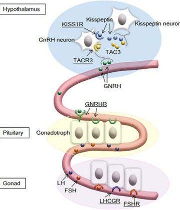
  • Known major G-protein–coupled receptors (GPCRs) and their ligands involved in the hypothalamus-pituitarygonadal axis. GPCRs are underlined. A gain-of-function mutation in KISS1R has been identified in a patient with precocious puberty.
Applications of GOF 
  • GOF studies are typically applied in virology and have revealed many details regarding the biological mechanisms responsible for viral transmission and replication. 
  • The high replication and mutation rates of viruses often lead to escape mutants. It is common for these new viral lineages to acquire genomic changes that reduce or eliminate the affinity of natural or vaccine-induced antibodies toward the virus. 
  • early studies regarding the E484K mutation of the severe acute respiratory syndrome coronavirus 2 (SARS-CoV-2) spike protein suggest affinity towards the angiotensinconverting enzyme 2 (ACE2) receptor, the target of the virus, is enhanced. Simultaneously, neutralization by serum antibodies sourced from patients who had previously recovered from wild-type SARS-CoV-2 infection is evaded more effectively. 
  • The U.S. Food and Drug Administration (FDA) requires animal testing on vaccines before human trials can begin. Since viral tropism towards the model species is unlikely to exist already, in cases where human viruses are under investigation, strains that can infect the model species must be generated. This can be achieved using GOFR, wherein the virus is passage through the animal, thereby allowing molecular determinants of transmissibility to be identified and vaccines under investigation to be tested.
SOFTWARE TOOLS FOR GENETIC MUTATION ANALYSIS 
  • Technological advancement has indeed contributed its quota in bioinformatics. Analysis of gene’s constituent for mutation can be done using software tools. 
  • The accuracy of these tools differs from one to the other. Here are some these tools considered in the review: Biomedical Mutation Analysis (BMA), MutaNET, VariantMaster, Mutation Surveyor, Mutation taster, Polyphen2, SIFT, KGGSeq, Galaxy, Ga TK, AlaMut, Imutant, SNPEffect, HOPE etc.

1.Biomedical Mutation Analysis (BMA) 
  • BMA was designed for an accessible analysis of mutations online. BMA is a user friendly application by which the user compare aligned sequences with a reference sequence. 
  • The output given will be the place where the changes are and to what it changed. Assists in computing changes in nucleotide and amino acid sequences.
  • BMA is an online application that stores all information related to the mutation analyses. This tool performs thanks to an analysis algorithm able to evaluate multiple patients, where each one can include multiple sequences.

SIGNIFICANCE AND PRACTICAL APPLICATION OF MUTATIONS 

Significance:- 
  • Mutations are the main source of variation in organisms of a species. 
  • Mutations provide a path for evolution of new varieties and/or species. 
  • Mutations have helped much in understanding the structure and function of gene.    Practical
Application:- 
  • Some mutations have beneficial effects and are useful in crop improvement. Mutations in both qualitative and quantitative traits have been exploited in plant breeding Examples:-
    1. Two amber grain coloured mutants viz., Sharabati sonara and 'Pusa lerma' have been produced in India from 'Sonara 64' and 'Lerma rajo' varieties of Mexican origin. 
    2. Using X-rays irradiation, semi dwarf varieties of rice have been produced. Till 1982 more than 45 useful varieties produced through mutation were in use. 
    3. Many more varieties in pulses, maize, Sorghum spices etc. have been produced through mutation breeding. 
    4. Early and late ripening varieties of wheat and rice have been developed by mutation. 
    5. Some varieties of maize, wheat, soyabean and other crops developed through mutation have been reported to be rich in protein and free amino acids like lysine.
  • Through mutations, resistance of various diseases have been raised in various crops. 
  • Mutation in a gene allow the gene to be studied in greater detail. 
  • Mutation is helpful in establishing the relationship between a gene and the protein produced by the gene. 
  • Biochemical mutations help in deciphering the biochemical pathway into individual reaction steps. Such a pathway can never be studied without mutational studies.

Disadvantage Of Mutation 
  • The process is generally random and unpredictable. 
  • Useful mutants recessive are rare and predominantly recessive. 
  • Mutants can have strong negative pleiotropic effects on other traits. 
  • Health risks: handling, chemical mutagens; radiations, fast neutrons treatments. 
  • Most mutants are of no use to breeding even if a large number of mutants can be produced.

REFERENCE
  1. Loewe, L., Genetic Mutation | Learn Science at Scitable. 2008,https://www.nature.com/scitable/ topicpage/geneticmutation-1127?error=cookies_not_supported&code= f4b6c104-de21-4785-987b-84d03ed7b087
  2. Brennan, J., How Can a Mutation in DNA Affect Protein Synthesis? 2018, https://sciencing.com/can-mutationdnaaffect-protein-synthesis-2028.ht
  3.  Khan S and Vihinen M., Performance of protein stability predictors. - PubMed - NCBI. 2019 https://www.ncbi.nlm.nih.gov/pubmed/20232415
  4. Genetics Home Reference, What is a gene mutation and how do mutations occur? 2019, https://ghr.nlm.nih.gov/primer/mutationsanddisorders/gen emutation 
  5. Bartee, L., How Gene Mutations Occur – Mt Hood Community College Biology 102. 2016, https://openoregon.pressbooks.pub/mhccbiology102/chapt, 

Regeneration of Plant in plant biotechnology

Contents

  • Introduction
  • History
  • Regeneration Of Plants
  • Types of regeneration in plant
  • Plant tissue culture
  • Types of tissue culture
  • Application of tissue culture
  • Advantages & Disadvantages of tissue culture
  • Conclusion
  • References 

Introduction

An entire plant can be regenerated from an adult tissue or organ, a mass of unorganized calli, or even a single cell in a process referred to as plant regeneration. Plant regeneration refers to the physiological renewal, repair, or replacement of tissue in plants. The totipotency or pluripotency of plant cells underlies the ability of plants to regenerate, reflecting the high plasticity of cell fate. Totipotency refers to the ability of a cell to differentiate into a complete individual, whereas pluripotency involves the differentiation of a specific group of tissues or organs from a cell. The concept of tissue culture was proposed as early as a century ago and envisaged the regeneration of whole plants from somatic cells in vitro. The tissue culture system has matured since the historical discovery that different concentration ratios of auxin and cytokinin (CK) are critical to regenerating adventitious roots and shoots. Steward et al. (1958) successfully regenerated new somatic embryos and subsequently developed roots and shoots by using isolated phloem cells from carrot roots, which confirmed the totipotency of plant cells. Since then, tissue culture technology based on regenerative ability has been extensively used in various fields, including basic research, micropropagation, and transgenic breeding. The ability of plant regeneration is affected by multiple factors, including use of a plant growth regulator, the composition of basic medium and explant type. Importantly, plant tissue culture presents strong species dependence and genotype specificity. Some plants, such as tobacco (Nicotiana tabacum), Arabidopsis thaliana, and rice (Oryza sativa), can be easily regenerated in vitro, whereas other plants, such as soybean (Glycine Max), wheat (Triticum aestivum), and maize (Zea mays), are more difficult to regenerate. Moreover, Japonica varieties show a higher capacity for callus formation than Indica varieties in rice. The tissue culture capacities of hybrid lines are higher than those of inbred lines in maize (Duncan et al., 1985). Clarifying the regulatory network and genetic control of plant-regeneration ability in tissue culture is helpful to improving plant-regeneration rates and genetic transformation efficiency.

  • Regeneration: - Regeneration is the natural process of replacing or restoring damaged or missing cells, tissues, organs, and even entire body parts to full function in plants and animals.
  • The process of growing an entire plant from a single cell or group of cells.
  • Regeneration is possible because plant cells can be made totipotent using hormones.
  • Differentiated tissue: stems, leaves, roots, etc.
  • Undifferentiated (embryonic) cells are totipotent can become a whole new plant by differentiating into a whole new.

History

  •  The theoretical basis for plant tissue culture was proposed by Gottlieb Haberlandt in his address to the German Academy of Science in 1902 on his experiments on the culture of single cells
  • Historically, Henri-Louis Duhamel du Monceau (1756) pioneered the experiments on wound healing in plants through spontaneous callus (unorganized mass of cells) formation on decorticated region of elm plants.
  • Vochting (1878) suggested the presence of polarity as a key feature that guide the development of plant fragments.
  •  In 1902, a German Botanist Gottlieb Haberlandt developed the concept of culture of isolated cells of Tradescantia in artificial condition. Though his experiment failed to induce the cells to divide.
  • Then the possibility for cultivation of plant tissues for unlimited period was announced simultaneously by P.R. White (1939) and R.J. Gautheret (1939).
  •  In 1959, discovery of kinetin promoted by F. Skoog along with C.O. Miller and co-workers and demonstration of induction of regeneration of shoots in tobacco callus paved the way for multiplication of plant by tissue culture.


THE PLANTS CAN BE REGENERATED BY: -


Organogenesis in plants

The process of development of plant organs such as shoot, flower, and root system from either an ex-plant or from the callus of culture is known as organogenesis in plants.

A completely developed plant consists of organs specialized in a particular function such as roots are responsible for absorbing nutrients and water from the soil, leaves are necessary for photosynthesis, and flowers for reproduction. Tissues such as meristem, cortex, phloem, and epidermis are organized together to form these organs. Developing and initiating these organs is called organogenesis.

 Meristematic cells are responsible for the development of plant organs like the root system, flowers, and shoot system. Shoot apical meristem or shortly known as SAM is responsible for generating or developing organs above the root, later organ. Shoot apical meristem (SAM) regenerated organs such as leaves, stems, buds, flowers, etc. hold organogenesis capability on their edges. When these cells are induced in-vitro a whole new plant grows from it. This whole process is called organogenesis.

Dedifferentiation and redifferentiation are the two steps that are involved in organogenesis.

Dedifferentiation is a process that helps in the formation of callus from the tissues of explant with acceleration in cell division. Cells multiply and divide very quickly to grow their number to form undifferentiated cells, i.e. callus, this process of dedifferentiation.

Redifferentiation is the process of developing a permanent organ by converting the cells that were formed during dedifferentiation. Cells lose their capability to multiply and divide in this process so that they can be converted into permanent tissue.

The process of organogenesis can take place in three ways:

  • From an explant
  • From the callus culture
  • From the axillary buds

Types of Organogenesis in plants

There are two types of organogenesis in plants that are:

  • Direct organogenesis
  • Indirect organogenesis

Direct organogenesis in plants tissue culture

When buds and shoots are directly developed from tissue and there is no need for the callus stage then this process is known as direct organogenesis in plant tissue culture.

Direct organogenesis results in the development of planting material with no genetic variation therefore cloning. Uniformity in the planting material is ensured. This process is also useful in propagating plants with a better multiplication rate (the number of plants per explant is higher).

Direct organogenesis is more of an industrial process as it provides plants with better multiplication rates and cloning propagation where the genetic variation is zero.

Indirect organogenesis in plant tissue culture

In this process of indirect organogenesis, a plant’s organ is developed from the callus of an explant (tissue that developed at the site of a cut or wound). The process of indirect organogenesis is more useful in the development of a transgenic plant. There are two ways that can be used to develop a transgenic plant in the indirect organogenesis method:

  • Transformed callus is used to regenerate a new plant that is transgenic
  • A modified explant is used to develop callus in the shoot, transform explant is initially used.

Factors that affect organogenesis:

We can divide the factors into two major groups that is:

  • External factors
  • Internal factors

External factors that affect organogenesis are

  • The medium

 The medium has a great impact on organogenesis

  •  The chemicals and medicines

Curtain chemicals such as auxin and cytokinin show a great impact on the growth of plant organs such as roots and shoots. Experiments and studies have shown that auxin simulates information of the root system and stops the formation of the shoot. Whereas, cytokinin promotes the development of shoot.

  • The environmental conditions

The environment and surroundings play a key role in organogenesis. A good and rich nutrient environment promotes organogenesis whereas a harsh and tough environment does the vice versa.

Internal factors that effects organogenesis is

  •  Gibberellin

Gibberellins is a hormone that restricts the formation of shoot and root both. Not only this but also gibberellins lower the content of starch and prohibit bud formation.

  •  Carbohydrates

Carbohydrates work as osmotic agents and as a respiratory energy source. Callus development and growth are affected by osmotic stress. Sucrose which is a form of carbohydrates is essential for this process.

  •  Ethylene

Ethylene is a hormone that enhances the development process during the primordial process but the process of organogenesis is blocked by ethylene hormones.

SOMATIC EMBRYOGENESIS

Somatic embryogenesis is the process wherein somatic cells differentiate into somatic embryos. It is not a naturally occurring process, an artificial one wherein an embryo or plant is obtained from one somatic cell. Somatic embryos take form from the cells of the plants, which usually do not take part in embryo development. Neither a seed coat nor endosperm is formed around the somatic embryo.

In the process, one cell or a cluster of cells initiates the developmental route, which results in reproducible regeneration of non-zygotic embryos, which can germinate for the formation of an entire plant.

The cells which are derived from potential source tissues are subject to a culture medium for the formation of an undifferentiated cluster of cells referred to as the callus. In the tissue culture medium, the plant growth regulators can be formed for the induction of the formation of calluses and hence modified to induce the embryos for the formation of calluses.

Process of Somatic Embryogenesis

The somatic embryogenesis procedure is a three-step procedure, which causes the induction of embryogenesis, development of the embryo and its maturation.

The principle of somatic embryogenesis finds its basis on the topic of totipotency of the plant cells; it illustrates two facets of plant embryogenesis:

  • The process of fertilization can be replaced by an endogenous mechanism.
  • The other types of cells of the plant, apart from the fertilized egg cells, can retrieve the capacity to form an embryo.

Since the process of somatic embryogenesis does not entail the procedure of fertilization, it promotes the large-scale propagation of plants at a faster rate. In addition, it also assists in the genetic transformation of plants, serving as a promising resource for the cryo-storage of the embryo and germplasm.

Somatic embryogenesis – Induction

Cells are reactivated to differentiate and develop embryos, which occur through two processes: direct somatic embryogenesis and indirect somatic embryogenesis.

Direct somatic embryogenesis

It involves the development of the embryos in a direct way from the cells of the explants, such as the cells of the immature embryos. Here, there is no intermediary stage (like the formation of the callus). The explants of the somatic embryogenesis are seen to entail PEDCs (pre-embryogenic determined cells).

Indirect somatic embryogenesis

It includes the formation of somatic embryos by reiterating numerous cycles of cell divisions. It includes intermediary steps of growth of the callus, and hence the process includes multiple steps.

The cells which do not carry the pre-embryogenic determined cells are caused to differentiate for the formation of the embryo by revealing different treatments. The cells modify into IEDs (induced embryogenic pre-determined cells).

Types of Somatic Embryogenesis

Somatic embryogenesis is of two types:

  • Direct somatic embryogenesis

Here, the embryos start directly from the explants when callus formation does not take place. Embryos, in this case, are formed as a result of Pre-induced Embryogenic Determined Cells (PEDCs).

  • Indirect somatic embryogenesis

The callus from the explants occurs from where the embryo develops. Here, the embryos are formed as a result of Induced Embryogenic Determined Cells (IEDCs).

Advantages of Somatic Embryogenesis

In comparison with zygotic embryogenesis, somatic embryogenesis has these benefits:

  • A huge number of embryos are obtained
  • The development and environmental stage of somatic embryos can be regulated
  • This process of embryogenesis can be monitored easily

The significance of somatic embryogenesis is as follows:

  • Production of artificial seeds
  • Higher rate of propagation
  • Apt in suspension culture
  • Labour savings

Factors Affecting Somatic Embryogenesis

The aspects which affect the process of somatic embryogenesis are as follows:

Traits of explant

Despite the fact that variations of explants can be used, the apt stage of development of explants is vital too to initiate the embryogenic callus; whereas juvenile explants tend to give rise to more somatic embryos compared to older explants. Also, different explant explants tissues from the same mother plant generated embryogenic callus at varying frequencies.

The desired species of plants to be induced for embryogenesis decides the choice of explants. For the majority of plant species, explants of immature zygotic embryos are apt for somatic embryogenesis.

Growth regulators

Cytokinins: -These have been in use in the primary medium consistently at the time of embryogenesis of the crop plants. They are vital in speeding up the process of maturation of somatic embryos, the cotyledon development, precisely.

Auxins: - These alone or in combination with cytokinin seemingly are vital for the start of growth and the induction of the embryogenesis of all the auxins. Auxins find immense importance in the first step of this process – the step of induction. High levels of auxins can lead to the inhibition of embryogenesis in the explants of the citrus plants.

Abscisic acid: -These are supplied at the inhibitory levels. It facilitates the development and maturation of the somatic embryos, while also inhibiting the unusual proliferation and the initiation of the accessory embryos.

Genotype

The process of embryogenesis is also affected by the genotypic variation seen in different plants; as per research, it can also be as a result of the endogenous levels of the hormones.

Sources of nitrogen

Nitrogen forms that are utilized in the media have an influence on the process of embryogenesis in plants. Forms of nitrogen have a marked influence on somatic embryogenesis. Somatic embryo development takes place on a medium that contains       NO3 as the only source of nitrogen.

Polyamines

The concentration of polyamines in media or explants is said to have an effect on the process. Experts observe the concentration of polyamines to be seen in higher concentrations in the polyembryonates compared to monoembryonates.

Electrical stimulation

Electrical stimuli apparently facilitate the differentiation of the structured embryo by influencing the cell polarity via modifications in the structure of the microtubules and the induction of first asymmetric division.


REGENERATION OF PLANTLETS

  1. Preparation of Suitable Nutrient Medium: Suitable nutrient medium as per objective of culture is prepared and transferred into suitable containers.
  2. Selection of Explants: Section of explants such as shoot tip should be done.
  3. Sterilization of Explants: Surface sterilization of the explants by disinfectants and then washing the explants with sterile distilled water is essential.
  4. Inoculation: Inoculation (transfer) of the explants into the suitable nutrient medium (which is sterilized by filter-sterilized to avoid microbial contamination) in culture vessels under sterile conditions is done
  5. Incubation: Growing the culture in the growth chamber or plant tissue culture room, having the appropriate physical condition (i.e, artificial light; 16 hours of photoperiod), temperature (-26°C) and relative humidity (50-60%) is required.
  6. Regeneration: Regeneration of plants from cultured plant tissues is carried out.
  7. Hardening: Hardening is gradual exposure of plantlets to an environmental condition.
  8. Plantlet Transfer: After hardening plantlets transferred to the green house or field conditions following acclimatization (hardening) of regenerated plants.

                                 Fig: - The schematic diagram of plant tissue culture       

Plant tissue culture

  • Plant tissue culture is a collection of techniques used to maintain or grow plant cells, tissues or organs under sterile conditions on a nutrient culture medium of known composition.
  • It is widely used to produce clones of a plant in a method known as micropropagation.
  • Plant tissue culture relies on the fact that many plant cells have the ability to regenerate a whole plant (Cellular totipotency)
  • single cells, plant cells without cell walls (protoplasts), pieces of leaves, stems or roots can often be used to generate a new plant on culture media given the required nutrients and plant hormones.
  •  Preparation of plant tissue for tissue culture is performed under aseptic conditions under HEPA filtered air provided by a laminar flow cabinet.

      Following are the main categories of Cultures: -

Primary Culture: - These model the natural function of the Tissue and are generally mortal. They consist of natural Tissues excised from the living organisms by biopsy.                                                         

Culture of Established Cell Lines: - These are derived from tumor biopsies, or from the primary cells that had undergone mutation and continued to replicate.

Some Types of Tissue Culture techniques

Seed Culture: In seed Culture, explants are obtained from an in- vitro derived plant and hence are introduced into a laboratory where they proliferate. To prevent the plants from Tissue damage it should be sterilized.

Embryo Culture: Embryo Culture involves the in-vitro development of an embryo. For this process, an embryo is isolated from and living organism, both a mature and an immature embryo can be used. Mature embryos can be obtained from ripe seeds whereas immature embryos are obtained from the seeds that failed to germinate. The ovule, seed, or fruit has already been sterilized, hence there is no need to sterilize them.

Callus Culture: - A callus can be defined as an unorganized, dividing mass of cells. A callus is the explants are Cultured in a proper medium good. The growth of callus is followed by organ differentiation. This Culture is grown on a gel-like medium composed of agar and specific nutrients which are required for the growth of the cells.

Organ Culture: - In organ Culture, any organ of the plant such as a shoot, the leaf can be used as an explant. Many methods can be used for the organ Culture such as the plasma clot method, raft method, the grid method, and Agar gel method. This method can be used to preserve the structure and functions of an organism

Meristem Culture: - meristems have the main function of the production of new cells and the synthesis of protoplasm. Shoot meristem consists of a group of certain actively dividing cells that are being protected by the developing leaves.

Protoplast Culture: - It can be defined as a cell without a cell wall. The hanging-drop method or micro-Culture chambers can be used to Culture a protoplast. A number of phases can be observed in protoplast Culture, development of cell walls, cell division, regeneration of a whole plant.

Suspension Culture: - suspension Culture can be defined as a form of Culture in which single cells or small aggregates of cells multiply while suspended in an agitated liquid medium. It can also be called cell Culture or cell suspension Culture.

Steps of Tissue Culture

  • Following are the steps of Tissue Culture

Initiation Phase: -

  • This is a stage when the Tissue is initiated into the Culture. To prevent the process from any contamination the Tissue of interest is obtained, introduced, and sterilized.

Multiplication Phase: -

  • In the multiplication stage, the sterilized ex-plant is introduced into the medium which consists of growth regulators and appropriate nutrients, they are responsible for the multiplication of cells. Hence this undifferentiated mass of cells is known as a callus.

Root Formation: -

  • This is the stage when the root starts forming. To initiate the formation of root plant growth hormones are added. Consequently, complete plantlets are obtained.

Shoot Formation: -

  •  For the formation of the shoot, plant growth hormones are added and growth is observed for a week.

Acclimatization: -

  • When the plant starts to develop, the plant is transferred to a greenhouse for it to develop under controlled environmental conditions. Thereafter it is finally transferred to the nurseries for its growth under natural environmental conditions.

HORMONES USED IN PLANT TISSUE CULTURE: -

Auxins: - Any of a group of plant hormones that regulate growth, particularly by stimulating cell elongation in stems.

Cytokinin's: - Cytokinin’s are a group of plant growth regulators which are primarily involved in performing cell division in plant roots, shoot system. 

Gibberellins: - One of the plant hormones that regulate a wide range of processes involved in plant growth, organ development, and environmental responses.

Abscisic Acid: -A plant growth regulator known for its functions, especially in seed maturation, seed dormancy, adaptive responses to biotic and abiotic stresses, and leaf and bud abscission.

Polyamines: - Polyamines can increase the activity of various antioxidant enzymes in plants, so that it can effectively regulate oxidative stress in plants caused by various environmental factors. 

Application of plant tissue culture

  • Plant tissue culture technology has been used in almost all the field of biosciences Its applications include
  • Production of phytopharmaceuticals and secondary metabolites.

        1.  Biotransformation (Biochemical Conversion)
        2.  Plant cell immobilization
        3.  Genetic transformation (Transgenic plant)
        4.  Elicitors

  • Micropropagation (Clonal Propagation)
  • Synthetic seed Protoplast culture and somatic hybridization
  • The influence of medium composition on alkaloid biosynthesis by Penicillium citrinum.
  • A Differential production of tropane alkaloids in hairy roots and in vitro culture
  • A Differential production of tropane alkaloids in hairy roots and in vitro cultured two accessions of Atropa belladonna L. under nitrate treatments.
  • Increased vincristine production from Agrobacterium tumefaciens C58 induced shooty teratomas of Catharanthus roseus G. Don.
  • Enhancement of taxane production in hairy root culture of Taxus x media var. Hicksii
  • Optimized nutrient medium for galanthamine production in Leucojum aestivum L. in vitro shoot system.
  • Extracts from black carrot tissue culture as potent anticancer agents.
  • Enhanced production of tropane alkaloids in transgenic Scopolia parviflora hairy root cultures over-expressing putrescine N-methyl transferase (PMT) and hyoscyamine-6B-hydroxylase (H6H).
  • Taxus globosa S. cell lines: initiation, selection and characterization in terms of growth, and of baccatin III and paclitaxel production.
  • Production of camptothecin in cultures of Chonemorpha grandiflora.
  • Regeneration, in vitro glycoalkaloids production and evaluation of bioactivity of callus methanolic extract of Solanum tuberosum L.


Conclusion: -

Plant regeneration is the major outcome of plant tissue culture, which is based on the principle of totipotency. Plant regeneration can be achieved by organogenesis and somatic embryogenesis. Organogenesis means formation of organs from the cultured explants. The shoot buds or monopolar structures are formed by manipulating the ratio of cytokinin to auxin in the cultures. In somatic embryogenesis, the totipotent cells may undergo embryogenic pathway to form somatic embryos, which are grown to regenerate whole plants. It was first established in carrots (Daucus carota), where bipolar embryos developed from single cells. The somatic embryogenesis is influenced by herbal extracts, phytohormones, and the physiological state of calli.

Plant regeneration involves the in vitro culture of cells, tissues, and organs under defined physical and chemical conditions. Critical for in vitro plant propagation and biotechnology, this phenomenon is also applicable to studies of plant developmental regulatory mechanisms.


References: -

  • D.Dumet, A. Adeyemi, O.B.Ojuederie (2008). Yam invitro genebanking. Genebank manual. http://www.iita.org/genebank/manual
  • D.Dumet, A.Adeyemi, O.B.Ojuederie (2008). Cassava in vitro processing and the genebanking. Genebank manual. http://www.iita.org/genebank/manual
  • HORT689/AGRO689 Biotechniques in Plant Breeding
  • H.S Chawla.2002 Introduction to Plant Biotechnology 2nd edition. Oxford & IBH Publishing C./ Pvt. Ltd New Delhi India
  • Murashige T. and Skoog F. (1962). A revised medium for rapid growth and bioassay with Tobacco tissue culture. Physiologia plantarum 15: 473-497.

Mendelian Inheritance Law of dominance, Law of Segregation, Law of independent assortment

Content Introduction History Who was Mendel Mendel’s Laws of Heredity Mendel’s Experiments The Rule of Unit Factors Gregor Mendel’s Disc...